| Parasite | Henneguya dogieli |
|---|---|
| Taxonomy | Myxozoa, Myxosporea, Bivalvulida |
| Hosts | Chinese perch (Siniperca chuatsi) |
| Infection site | Gill |
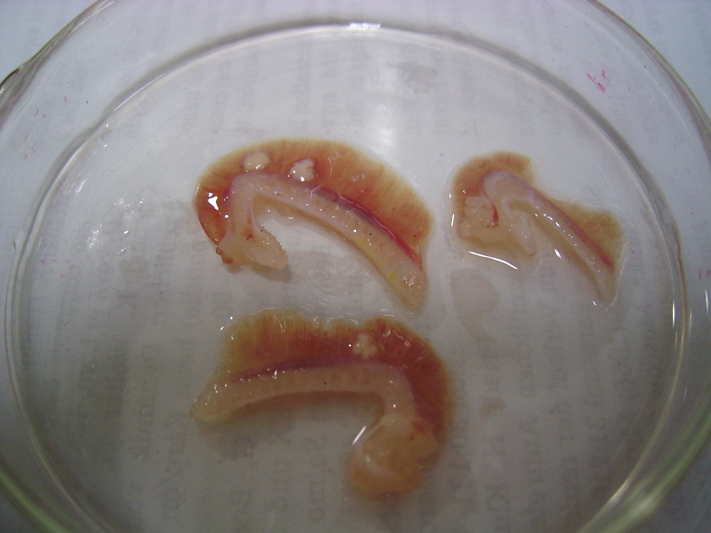
| Clinical signs | Lobate (flower-like) cysts are observed in the gill (Fig. 1). |
| Parasitology | A number of spores are formed inside the cyst (Fig. 2). A spore (length 12.8 (10.8-13.2) mm; width 7.0 (6.2-7.2) mm; thickness 5.8 (5.4-6.2) mm; length of caudal appendage 44.6 (15.6-54.0) mm) is fusiform and has 2 equal polar capsules (length 4.9 (4.8-5.0) mm; width 2.4 (2.0-2.6) mm) (Chen and Ma, 1998). The life cycle is unknown. |
| Pathology | Unknown |
| Health hazard | Since this parasite is not infectious to human, it is harmless in food hygiene. |
| Diagnosis | Check the spores by the wet-mount of squashed cysts. Sample should be smeared and stained by Giemsa or Diff-Quik. |
| References | Chen, Q. L., and C. L. Ma (1998): Myxozoa, Myxosporea. Fauna Sinica. Beijing, Science Press, 987 p. |
Fig. 2. Fresh spores of Henneguya dogieli
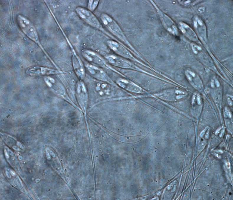

(Photos by Jin-Yong Zhang)
Fig. 1. Lobate cysts are observed in the gill of Chinese perch.